

by Filippo Cavallaro, PT; Simona Portaro, MD, PhD; Teresa Pintaudi, PT; Mariachiara Ceccio, PT; and Angelo Alito, MD
All authors are with the University of Messina in Messina, Italy.
Funding: No funding was provided for this article.
Disclosures: The authors have no conflicts of interest relevant to the content of this article.
Innov Clin Neurosci. 2023;20(1–3):10–12.
Abstract
Facial nerve palsy is a clinical diagnosis differentiating between central upper motor neuron lesions and peripheral lower motor neuron lesions. Rehabilitation is an important issue in peripheral facial nerve palsy management. In this article, we present the case of an adult woman affected by right peripheral facial nerve palsy due to acoustic neuroma surgical excision. She immediately started a rehabilitation plan, but it was stopped due to COVID-19 lockdown and did not resume because of the fear of contracting severe acute respiratory syndrome coronavirus 2 (SARS-CoV-2). Therefore, we planned to treat her palsy with remote neurocognitive rehabilitation. After 10 months of treatment, the patient underwent a follow-up physiatric assessment, confirming right facial palsy improvement. There was a slight nasolabial groove flattening and slight left oral rime deviation while smiling (House-Brackmann classification improved from Grade IV to III). Telerehabilitation represents a valid strategy for neurocognitive rehabilitation, not only in a pandemic scenario, but also in other conditions that lead to social distancing.
Keywords: Facial nerve palsy, acoustic neuroma, telerehabilitation, cognitive therapeutic exercise, rehabilitation, COVID-19
Facial nerve palsy is a clinical diagnosis differentiating between central upper motor neuron lesions (e.g., stroke) and peripheral lower motor neuron lesions (e.g., idiopathic or caused by infection or trauma or surgery).1–4 Peripheral facial nerve palsy has various causes; the most common is Bell’s palsy (approximately 75% of all cases), which has a better prognosis than secondary causes. Secondary causes include neoplasms, such as acoustic neuroma and facial nerve schwannoma, and/or surgical adverse effects.1–4 It has been reported that after removal of an acoustic nerve schwannoma or neuroma, facial nerve palsy might occur in up to 70 percent of cases.5 Rehabilitation is an important issue in peripheral facial nerve palsy management. Different approaches have been applied, such as exercise therapy, electrotherapy, massage, lymph drainage, and biofeedback therapy,6 but there is no evidence that any particular technique is better than others.7
Case Presentation
Herein, we present the case of an adult woman affected by right peripheral facial nerve palsy due to acoustic neuroma surgical excision two months before visiting our institution. During this time, she took steroids and neurotrophic drugs, with poor beneficial effects.
On admission, at the first physiatric assessment, she presented with a peripheral right facial nerve palsy with positive Bell’s and Nigro’s signs, right lagophthalmos, complete nasolabial groove, and right frontal wrinkles smoothing (House-Brackmann Grade IV).8 Therefore, a rehabilitation plan was immediately started but, two days later, it was stopped due to the COVID-19 lockdown. Thus, in order to guarantee an early and proper rehabilitation plan, we decided to treat her palsy with a remote rehabilitation approach via WhatsApp (Meta; Menlo Park, California, United States).
The patient was informed about the scope and procedures of the study per the Declaration of Helsinki, and written, informed consent for participation in the rehabilitation protocol was obtained and sent via email to the healthcare team.
Treatment
A smartphone application that could be utilized as a costless telerehabilitation system was used to reach rehabilitation goals (i.e., motor outcomes). Telerehabilitation treatment lasted 10 months in an asynchronous modality, with the healthcare team sharing texts, images, and video messages containing exercise instructions (timing and modalities), giving the patient time to understand how to perform them. Since it has been shown that emotional and communicative disorders can develop in patients with facial nerve palsy, we integrated psychological interventions into the treatment plan as well.9,10
The therapeutic plan included three phases. The first phase was based on a neurocognitive approach. The physiotherapist sent vocal message task instructions twice per week, using photos representing emotional facial expressions.11 In each session, the patient had to stay in a quiet, distraction-free environment and record and send the session to the rehabilitation team. Before starting self-treatment, she had to focus on images, representing happiness, sadness, interest, disgust, fear, anger, and surprise, for a few minutes, and then bring them to mind, paying attention to all the details she considered important. Next, she had to mentally represent those facial expressions with closed eyes, then reproduce them. The second phase was performed with a trained caregiver help (patient’s father), who had to manually stimulate soft facial tissues and mimic muscles. The third phase consisted of investigating and promoting sensory perception through recognition of different textures, sizes, and surfaces (e.g., recognizing rulers or brushes using lip and skin contact) while using cognitive sensory motor training therapy.
Psychological support was provided once per month or as needed. The Beck Depression Inventory12 was applied to monitor the patient’s psychological condition, but no signs of depression emerged. This approach not only helped the patient with her facial nerve palsy, but also helped manage her fear of COVID-19. It has been shown that self-competence in emotional regulation during the COVID-19 pandemic was not sufficient to cope with the trauma of the pandemic. The psychological support provided may have had an effect on rumination and worries, leading to improved self-confidence.13 After the lockdown ended, we repeatedly asked the patient to continue the rehabilitation protocol as hospital outpatient treatment, but she refused, preferring remote rehabilitation because of a fear of contracting COVID-19.
Outcome and Follow-up
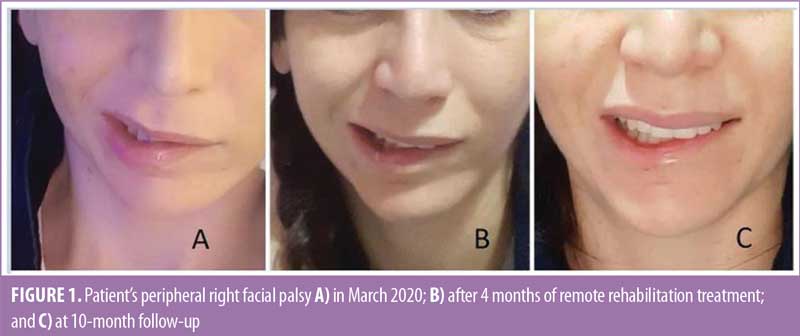
After 10 months of treatment, the patient underwent a follow-up physiatric assessment, confirming right facial palsy improvement (Figure 1). There was a slight nasolabial groove flattening and slight left oral rime deviation while smiling; House-Brackmann classification thus improved from Grade IV to III.8 Moreover, the patient expressed feeling comfortable and cared for by the rehabilitation team, even during the pandemic scenario. Although this remote rehabilitation approach was applied as an emergency option, this method proved to be helpful from both a clinical and psychological perspective.
Discussion
To the best of our knowledge, this is the first report determining the effects of a neurocognitive approach through mirror therapy and motor imagery via WhatsApp, without the reported, integrative, traditional rehabilitative method to treat iatrogenic peripheral facial palsy.14
Acoustic neuroma, also known as a vestibular schwannoma, is a slowly growing benign tumor typically originating from the vestibular nerve and represents the most common cerebellopontine angle lesion. Treatments include surgery with or without stereotactic radiotherapy.15
During surgery, it is mandatory to accurately identify the facial nerve course to preserve its function, avoiding skull base surgery complications.16 Management largely depends on nerve integrity. Consequences of this injury can be devastating and include peripheral facial palsy, exposure keratosis and blindness, disfigurement, poor nasal airflow, oral incompetence, synkinesis, and psychological stress.17 Moreover, facial nerve injuries alter facial shape, which can have a significant impact on an individual’s social life.
It is important to promptly identify and treat patients at high risk of poor long-term outcomes to reduce persistent facial nerve dysfunction risk and psychological distress.18 Various treatments to support recovery from this condition, such as mime therapy, electrostimulation, acupuncture, mirror therapy, and biofeedback, have been proposed.19 Mirror therapy has been reported as a good method to treat neurological conditions,20,21 with better results when preceded by motor imagery to allow the patient to consciously perceive a movement.14,22
However, to date, no studies suggest that one approach is more effective than the others. This lack of guidelines and specific protocols, along with the social distancing imposed by the COVID-19 lockdown, led us to develop a telerehabilitation approach. Recently, it has been shown that telerehabilitation is a valid option that should be adapted as soon as possible to face the COVID-19 crisis. This approach offers positive clinical results that are comparable, in some cases, to conventional face-to-face rehabilitation.23
Limitations. This case report has several limitations due to the lack of a recognized telerehabilitation device, but as an emergency option, it was the only way to guarantee rehabilitation strategies for our patient.
Conclusion
In conclusion, our telerehabilitation approach helped our patient, thus presenting as a valid strategy when social distancing is imposed, such as in pandemic scenarios or other situations (e.g., immunocompromised individuals).
References
- Finsterer J. Management of peripheral facial nerve palsy. Eur Arch Otorhinolaryngol. 2008;265(7):743–752.
- Peitersen E. Bell’s palsy: the spontaneous course of 2,500 peripheral facial nerve palsies of different etiologies. Acta Otolaryngol Suppl. 2002;(549):4–30.
- Roob G, Fazekas F, Hartung HP. Peripheral facial palsy: etiology, diagnosis and treatment. Eur Neurol. 1999;41(1):3–9.
- Ryzenman JM, Pensak ML, Tew JM Jr. Facial paralysis and surgical rehabilitation: a quality of life analysis in a cohort of 1,595 patients after acoustic neuroma surgery. Otol Neurotol. 2005;26(3):516–521.
- Wilson CM, Ronan SL. Rehabilitation postfacial reanimation surgery after removal of acoustic neuroma: a case study. J Neurol Phys Ther. 2010;34(1):41–49.
- Azuma T, Nakamura K, Takahashi M, et al. Electroneurography in the acute stage of facial palsy as a predictive factor for the development of facial synkinesis sequela. Auris Nasus Larynx. 2018;45(4):728–731.
- Pourmomeny AA, Asadi S. Management of synkinesis and asymmetry in facial nerve palsy: a review article. Iran J Otorhinolaryngol. 2014;26(77):251–256
- House JW, Brackmann DE. Facial nerve grading system. Otolaryngol Head Neck Surg. 1985;93(2):146–147.
- van Landingham SW, Diels J, Lucarelli MJ. Physical therapy for facial nerve palsy: applications for the physician. Curr Opin Ophthalmol. 2018;29(5):469–475.
- Beurskens CH, Heymans PG, Oostendorp RA. Stability of benefits of mime therapy in sequelae of facial nerve paresis during a 1-year period. Otol Neurotol. 2006;27(7):1037–1042.
- Osgood CE. Dimensionality of the semantic space for communication via facial expressions. Scand J Psychol. 1966;7(1):1–30.
- Beck AT, Steer RA, Brown GK. BD-II: Beck Depression Inventory Manual, 2nd edition. Psychological Corp; 1996:82.
- Carey TA, Griffiths R, Dixon JE, Hines S. Identifying functional mechanisms in psychotherapy: a scoping systematic review. Front Psychiatry. 2020;11:291.
- Paolucci T, Cardarola A, Colonnelli P, et al. Give me a kiss! An integrative rehabilitative training program with motor imagery and mirror therapy for recovery of facial palsy. Eur J Phys Rehabil Med. 2020;56(1):58–67.
- Wright A, Bradford R. Management of acoustic neuroma [published correction appears in BMJ. 1995;311(7017):1421]. BMJ. 1995;311(7013):1141–1144.
- Samii M, Matthies C. Management of 1000 vestibular schwannomas (acoustic neuromas): the facial nerve–preservation and restitution of function. Neurosurgery. 1997;40(4):684–695.
- Melvin TA, Limb CJ. Overview of facial paralysis: current concepts. Facial Plast Surg. 2008;24(2):155–163.
- Huang B, Xu S, Xiong J, et al. Psychological factors are closely associated with the Bell’s palsy: a case-control study. J Huazhong Univ Sci Technolog Med Sci. 2012;32(2):272–279.
- Agostini F, Mangone M, Santilli V, et al. Idiopathic facial palsy: umbrella review of systematic reviews and meta-analyses. J Biol Regul Homeost Agents. 2020;34(4):1245–1255.
- Zangrando F, Paolucci T, Vulpiani MC, et al. Chronic pain and motor imagery: a rehabilitative experience in a case report. Eur J Phys Rehabil Med. 2014;50(1):67–72.
- Ramachandran VS, Altschuler EL. The use of visual feedback, in particular mirror visual feedback, in restoring brain function. Brain. 2009;132(Pt 7):1693–1710.
- MacIntyre TE, Madan CR, Moran AP, et al. Motor imagery, performance and motor rehabilitation. Prog Brain Res. 2018;240:141–159.
- Suso-Martí L, La Touche R, Herranz-Gómez A, et al. Effectiveness of telerehabilitation in physical therapist practice: an umbrella and mapping review with meta-meta-analysis. Phys Ther. 2021;101(5):pzab075.